March 14, 2023 | RCE.ai
Significant developments and evolution of medical device technology have occurred in many fields of medicine. One such area was radiology, and studying history helps us understand how the past shaped the medical industry’s trajectory from conception to impact in patient care.
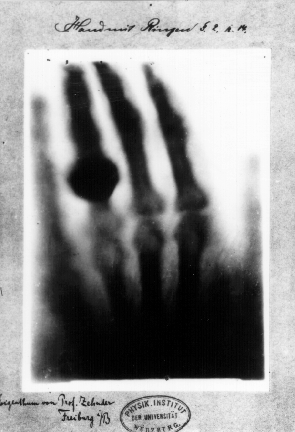
Back in 1895, German physics professor Wilhelm Röntgen discovered x-rays, and its transformational application in medicine to look within human tissue. The field of radiology advanced to leverage this capability to identify broken bones, pneumonia, scoliosis, cancerous nodules in bones, breast cancer, teeth abnormalities, amongst others.
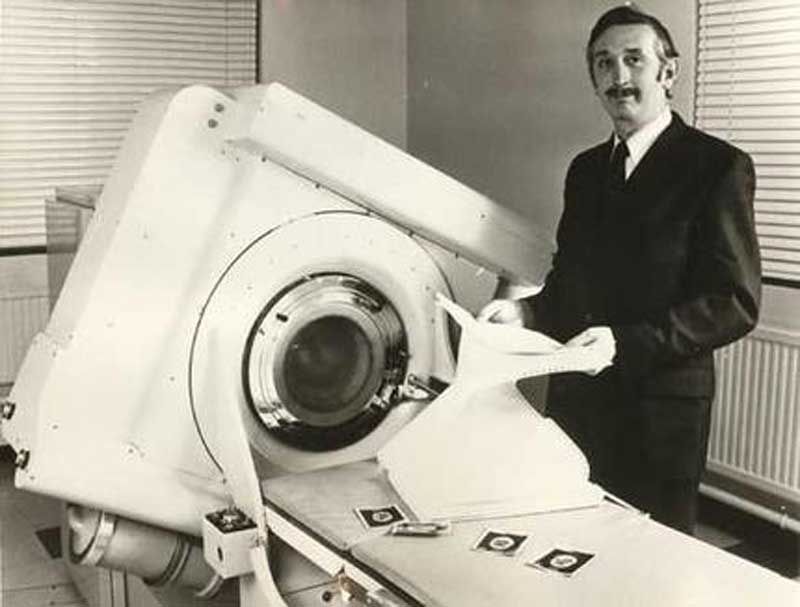
Few decades later, British engineer Godfrey Hounsfield and physicist Dr. Allan Cormack invented the ct-scan technology that combines a series of X-ray images taken from different angles around the body to create cross-sectional images of the bones, blood vessels and soft tissues inside your body. These images provide more detailed information than plain X-rays, similar to a loaf of bread providing more information about the bread, rather than just a slice of bread.
Fast forward many decades, we now have handheld technologies like OXOS that could revolutionize the way radiology can empower point-of-care imaging and diagnostics for clinicians and patients. These optimize the hospital-based C-Arm, a mobile imaging technique for fluoroscopic imaging during surgical and orthopedic procedures.
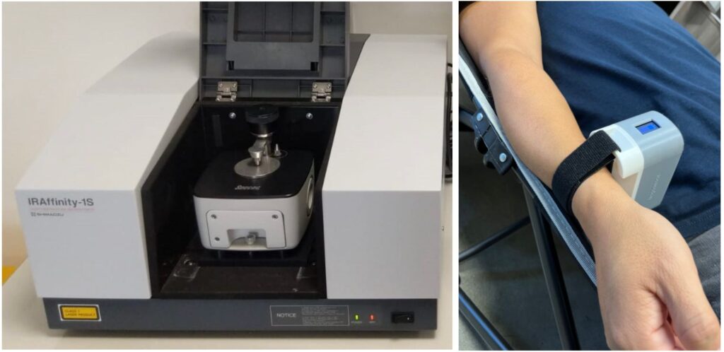
Another transformational technology is Infrared Spectroscopy. In specific, Fourier Transform Infrared Spectroscopy (FTIR), a technique that is used to obtain an infrared spectrum of absorption of an analyte of interest. Widely used in the fields of geology, chemistry, materials and biology research, the FTIR collects high-resolution spectral data over a wide spectral range, providing useful characteristic information about the analyte. The analyte could be a protein, an enzyme, or a cell signal in times of distress such as in heart attacks. Our team of top scientists, researchers and clinicians have collaborated to invent the wrist-worn transdermal infra-red spectrophotometric sensor (Transdermal-ISS) that probes the underside of the wrist providing similar selectivity as the FTIR spectrometer, without the need for a sample specimen.
We often get asked what we do, here at RCE?
And, we often think of a similar analogy that transitioned from the conventional x-ray to ct-scan, and subsequently point of care and mobile imaging. While based on similar underlying physics involving electromagnetic radiation, innovation via medical device provides the versatily to provide deeper information of the underlying anatomical or pathophysiological state of the patient. This has the potential to empower healthcare providers in providing better care for cardiac patients.